Open Snake Tracing System
From FarsightWiki
(Difference between revisions)
| (24 intermediate revisions by one user not shown) | |||
| Line 1: | Line 1: | ||
Tracing System (under construction) | Tracing System (under construction) | ||
| − | + | The code for the basic 3-D open-curve tracing system is now in the SVN. The latest code containing the 4-D open-curve tracing model, microglia processing code, GPU implementations and segmentation code will be released soon. We are working on the user manual for the software system. | |
| − | + | ||
| − | + | ||
| − | + | ||
| − | [[Image: | + | ---- |
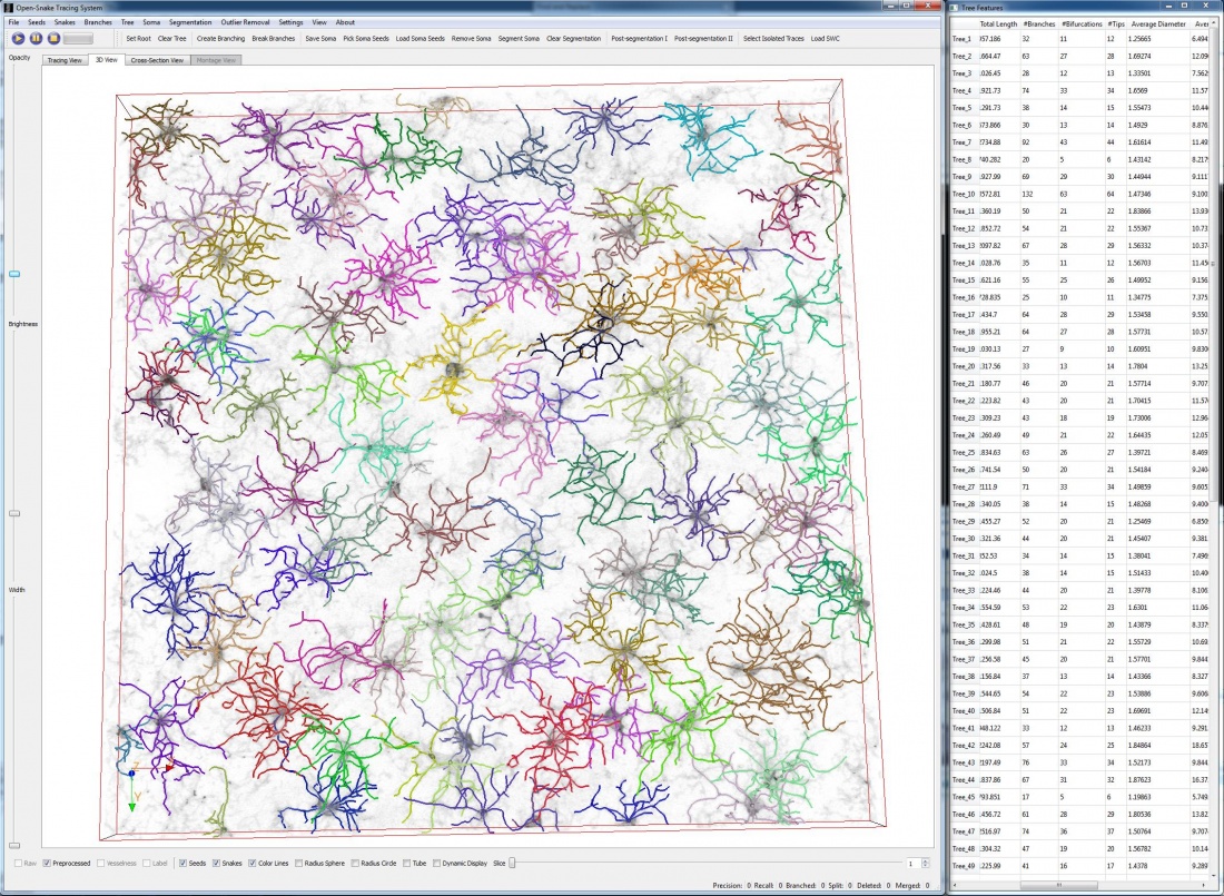
| + | [[Image:Screenshot2.jpg|thumb|center|1100px| Figure 1: ScreenShot of the Tracing System. 87 microglia cells were traced automatically and their identities are color-coded in the rendering. The feature table shows a set of features for each cell. ]] | ||
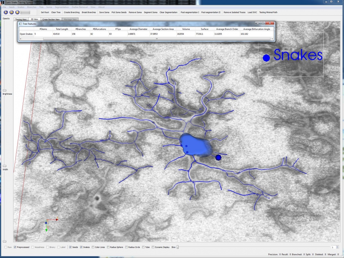
| + | [[Image:Screenshot1.jpg|thumb|center|1100px| Figure 2: ScreenShot of the Tracing System. Close-up view of one microglia cell, the soma/nuclei is segmented automatically.]] | ||
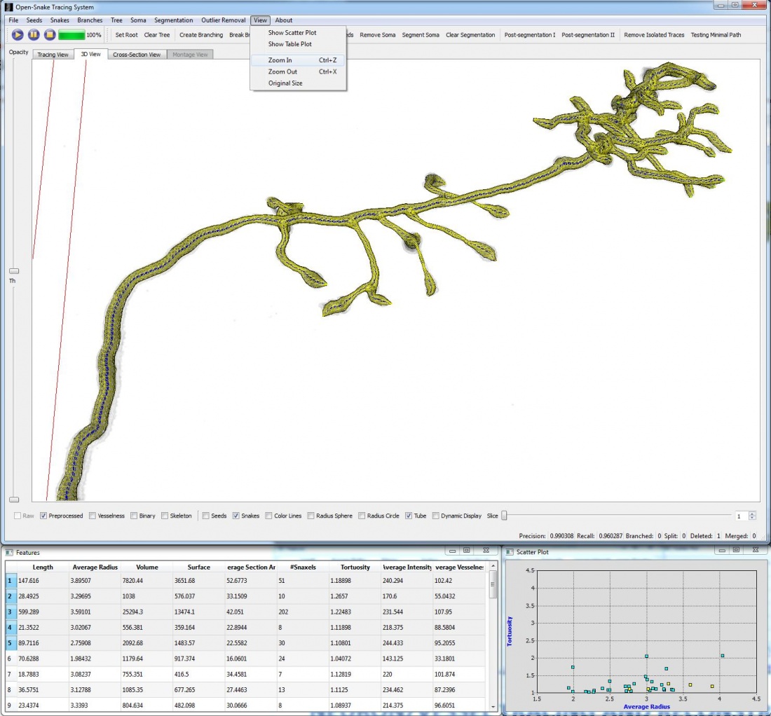
| + | [[Image:Screenshot.jpg|thumb|center|1100px| Figure 3: ScreenShot of the Tracing System. Tracing result was produced with 4-D open-curve snake model, which traces the centerline and estimates the radius simultaneously. ]] | ||
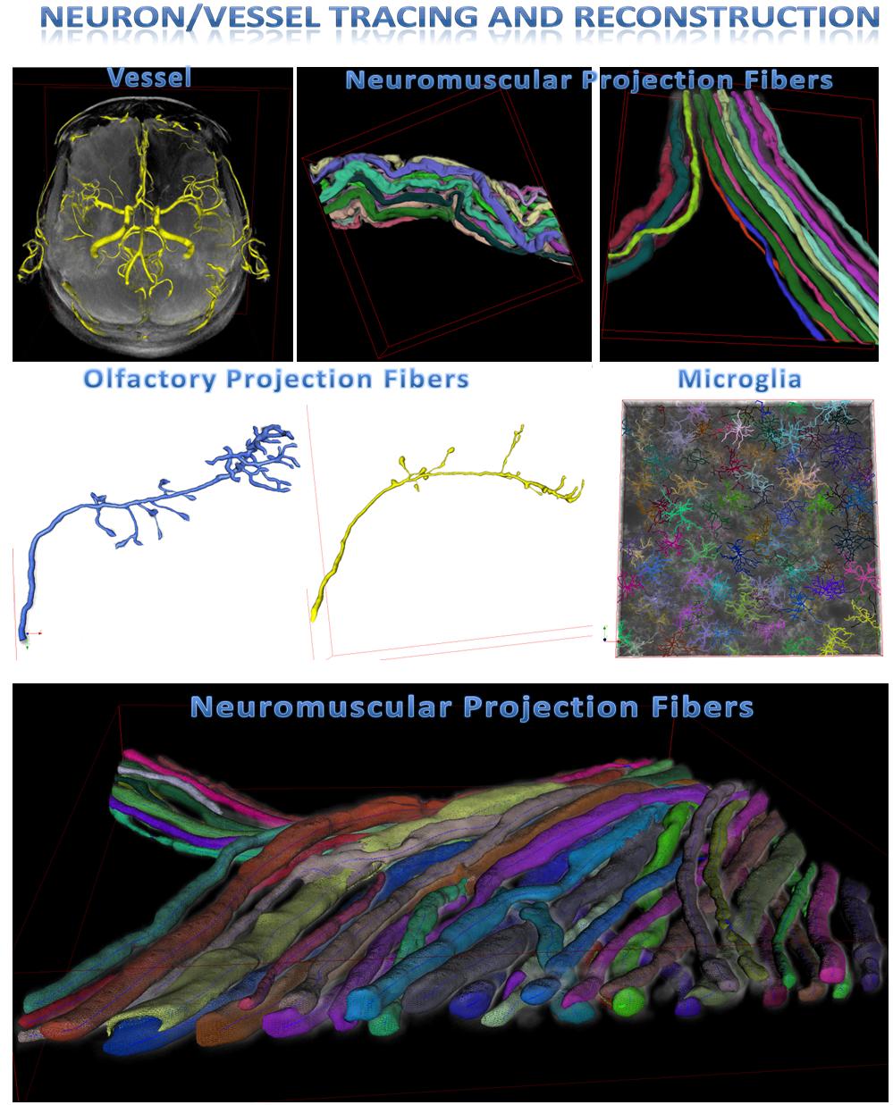
| + | [[Image:Results.jpg|thumb|center|1100px| Figure 4: Tracing and Reconstruction Results of Our System on Different Datasets]] | ||
Latest revision as of 16:46, 10 May 2011
Tracing System (under construction)
The code for the basic 3-D open-curve tracing system is now in the SVN. The latest code containing the 4-D open-curve tracing model, microglia processing code, GPU implementations and segmentation code will be released soon. We are working on the user manual for the software system.
- Contributors to the Tracing System:
- Yu Wang [1]
- Arunachalam Narayanaswamy
- Charlene Tsai


