File:Hippocampus.jpg
From FarsightWiki
Size of this preview: 800 × 374 pixels
Full resolution (972 × 455 pixels, file size: 257 KB, MIME type: image/jpeg)
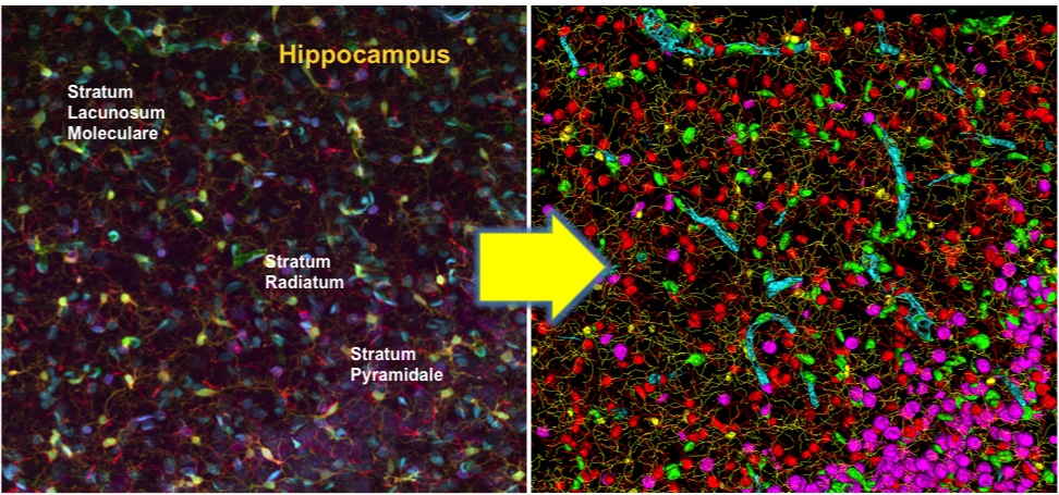
Cytovascular brain tissue mapping example. On the left is the maximum-intensity projection of a rat hippocampus imaged by multi-spectral confocal microscopy, and spectral unmixing to produce 5 channels. (Cyan) CyQuant-labeled cell nuclei; (Purple) NeuroTrace-labeled Nissl substance; (yellow) Iba1-labeled microglia; (red) GFAP-labeled astrocytes; and (green) EBA-labeled blood vessels. On the right is a composite frontal 3-D rendering of the multi-channel segmentation and cell classification results.
File history
Click on a date/time to view the file as it appeared at that time.
| Date/Time | Thumbnail | Dimensions | User | Comment | |
|---|---|---|---|---|---|
| current | 20:00, 29 April 2009 | | 972×455 (257 KB) | Roysam (Talk | contribs) | |
| 15:51, 22 April 2009 | 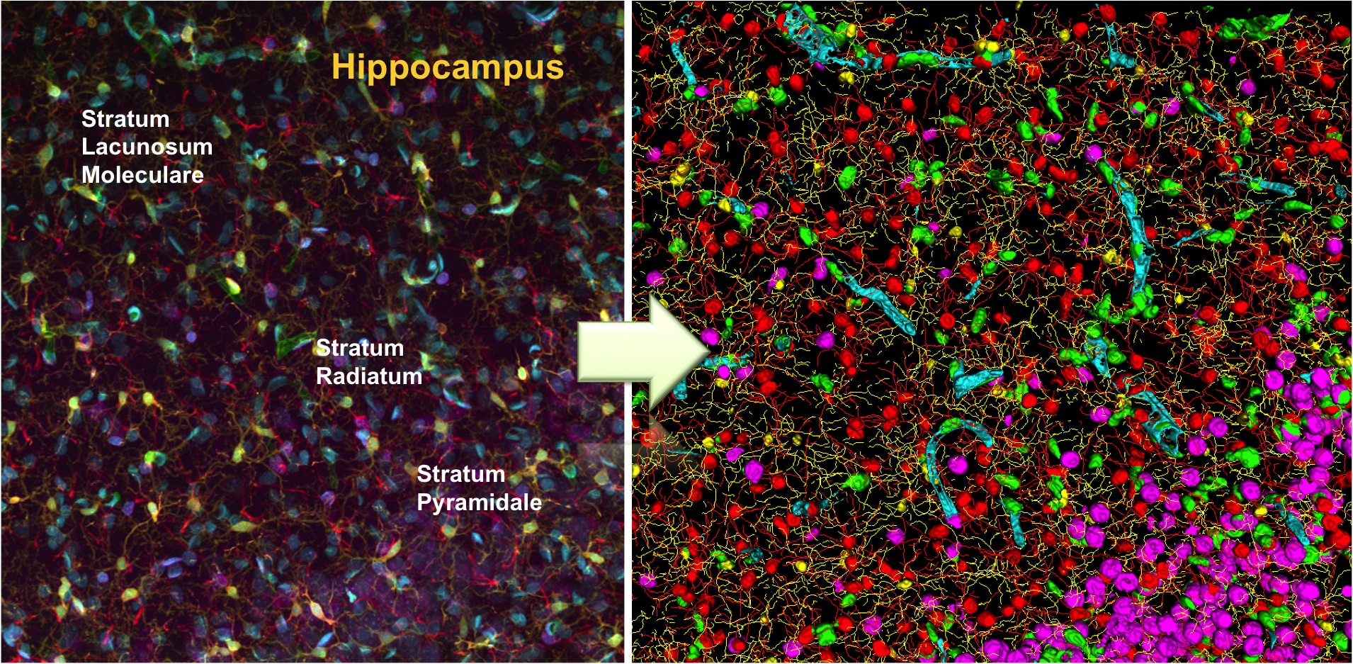 | 1,910×937 (1.14 MB) | Roysam (Talk | contribs) | (Cytovascular brain tissue mapping example. On the left is the maximum-intensity projection of a rat hippocampus imaged by multi-spectral confocal microscopy, and spectral unmixing to produce 5 channels. (Cyan) CyQuant-labeled cell nuclei; (Purple) NeuroTr) |
- Edit this file using an external application (See the setup instructions for more information)
File links
The following page links to this file: